

What is a tibial tubercle distalising osteotomy?
A tibial tubercle distalising osteotomy is an operation to lower a kneecap (patella) that sits too high, a condition called patella alta. The tibial tubercle is the bony bump at the top of the shin bone (tibia) where the patellar tendon attaches. The kneecap hangs from this tendon, so moving the tubercle further down the shin bone brings the kneecap down with it. A high-riding kneecap enters its groove at the end of the thigh bone later as the knee bends, which makes it easier for it to slip out. Patella alta is one of the main anatomical risk factors for kneecap dislocation (PMID 27077588; PMID 36728086).
Through an incision at the front of the upper shin, the surgeon cuts the tubercle free from the shin bone (an osteotomy), moves it down by a planned distance and fixes it with screws while the bone heals. In published studies the tubercle was moved down by an average of about 9 to 12 mm (PMID 34038256; PMID 33553451). It is often also moved towards the inner side of the knee, as in a tibial tubercle medialising osteotomy, and the operation is commonly combined with reconstruction of the medial patellofemoral ligament (MPFL), the main ligament that holds the kneecap on the inner side (PMID 34038256; PMID 35438591). It is one of several operations used for patella stabilisation.
Modern imaging is central to the decision. Kneecap height is measured on a side-on X-ray or MRI with ratios such as the Caton-Deschamps index, which compares the length of the joint surface of the kneecap with its distance from the top of the shin bone. CT or MRI scans also show other contributing factors, such as the position of the tubercle and the shape of the groove (PMID 27077588).
What are the benefits of a tibial tubercle distalising osteotomy?
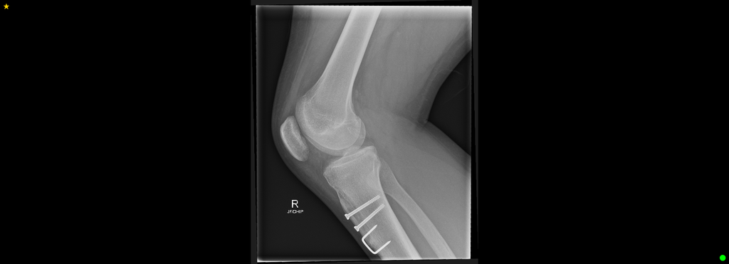
The main benefit is that the kneecap is brought back to a more normal height, so that it enters its groove earlier as the knee bends.
The kneecap becomes more stable. In one series, 95.6% of patients had no further dislocation (PMID 35438591), and a review of treatments for patella alta found that tibial tubercle distalisation was generally successful in preventing recurrent dislocation (PMID 23392290). In another study, the feeling that the kneecap might slip out (apprehension) resolved in all patients (PMID 33553451).
Knee function and pain also improve. In a review of 546 knees treated surgically for kneecap instability with patella alta, knee function scores improved by an average of about 32 points (PMID 33997063), and in one study pain at the front of the knee resolved completely in 71% of patients (PMID 33553451).
Correcting kneecap height also removes one of the main risk factors for failure of a soft-tissue stabilisation such as MPFL reconstruction (PMID 39763469; PMID 34038256).
What are the risks of a tibial tubercle distalising osteotomy?
To move the tubercle downwards, it has to be completely detached from the shin bone (PMID 40045470). Osteotomies with complete detachment of the tubercle have had a higher complication rate than those in which it is left partly attached (PMID 25980400). A review of 420 knees found complications in 7.6% and further operations in 14.3%, mostly to remove screws (PMID 34038256), and procedures on the tubercle carry a higher risk of major complications and further surgery than soft-tissue procedures alone (PMID 33997063).
Stiffness from scar tissue in the knee (arthrofibrosis) is one of the most common complications. In one series it affected 8.8% of patients and was more likely when the tubercle was moved further. The same series had fractures of the shin bone in 4.4% (PMID 35438591). Delayed healing or failure of the bone to heal (non-union) and infection can also occur (PMID 38516864; PMID 38371997). In patients younger than 21, complications occurred in 27% after distalisation, most commonly infection and stiffness (PMID 40045470).
Kneecap height may drift up again over time. In one study, 88% of patients who had X-rays at least a year after surgery again measured as patella alta, although the clinical importance of this is not yet known (PMID 40949532). The kneecap can still feel unstable, with apprehension remaining in 15 to 33% of patients in some studies (PMID 23392290).
Distalisation is not always needed. In one study of people with a high kneecap and a tubercle positioned too far to the outer side, adding distalisation to medialisation gave no better results (PMID 36057569). There has also been concern that lowering the kneecap could increase pressure on the cartilage behind it and cause pain. In one study, persistent pain after surgery was linked with the severity of cartilage damage at the time of surgery (PMID 33553451). Other risks are those of knee surgery in general, such as blood clots and numbness around the incision.
Summary
A tibial tubercle distalising osteotomy moves the bony attachment of the patellar tendon further down the shin bone to lower a high-riding kneecap, so that it sits more securely in its groove and is less likely to dislocate. It is mainly used when imaging shows patella alta in people with recurrent kneecap dislocation, often together with an MPFL reconstruction or medialisation of the tubercle. It is usually recommended when the expected benefits for you outweigh the risks. You should discuss your symptoms, your scans and your goals, including plans for sport and work, with your surgeon so that you can consider the options and indications for an osteotomy together.
General information only. The information on this page is general in nature and does not replace individual medical advice. All surgery carries risks, and results vary between patients. Please discuss your own circumstances with your treating practitioner.