
What is a tibial tubercle medialising osteotomy?
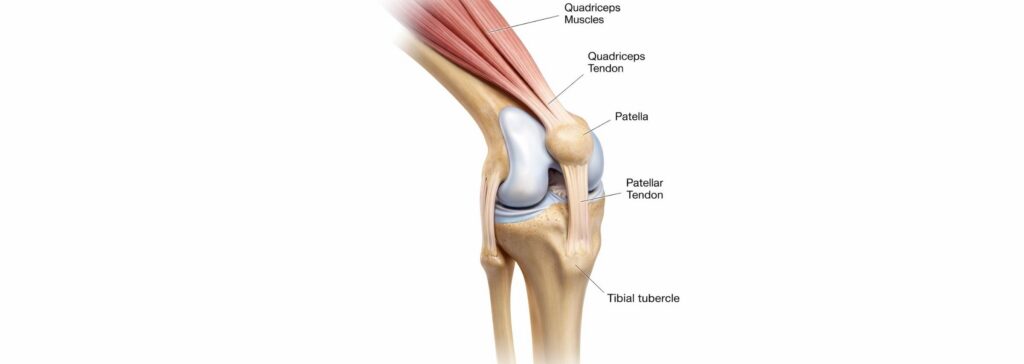
A tibial tubercle medialising osteotomy is an operation to improve the stability and tracking of the kneecap (patella). The tibial tubercle is the bony bump at the top of the shin bone (tibia) where the patellar tendon attaches. The patellar tendon works like a rope that links the kneecap to the shin bone, so the position of the tubercle sets the direction in which the kneecap is pulled when the thigh muscles contract. In some people the tubercle sits too far towards the outer side of the knee. This pulls the kneecap outwards and can contribute to it dislocating (PMID 36728086).
Through an incision at the front of the upper shin, the surgeon makes a controlled cut in the bone (an osteotomy), moves the tubercle a planned distance towards the inner side of the knee (medialisation) and fixes it with screws while the bone heals. In the Elmslie-Trillat technique the tubercle is moved straight inwards. In the Fulkerson technique (anteromedialisation) it is moved inwards and slightly forwards (PMID 25980400; PMID 24197613).
The operation is used to treat recurrent kneecap dislocation and, in the anteromedialisation form, wear of the cartilage on the back of the kneecap (PMID 24197613; PMID 41207749). Moving the tubercle forwards as well as inwards reduces the pressure on the outer part of the kneecap (PMID 36453729). It is one of several operations used for patella stabilisation.
Modern imaging is central to the decision. CT or MRI scans measure the distance between the tibial tubercle and the groove at the end of the thigh bone in which the kneecap runs (the TT-TG distance), and show other contributing factors such as a high-riding kneecap or a shallow groove (PMID 36728086). The osteotomy is often combined with reconstruction of the medial patellofemoral ligament (MPFL), the main ligament that holds the kneecap on the inner side (PMID 39465420). A knee arthroscopy may be performed at the same time to confirm the condition of the cartilage (PMID 23539699).
What are the benefits of a tibial tubercle medialising osteotomy?
The main benefit is a more stable kneecap. By moving the attachment of the patellar tendon, the operation corrects the line of pull on the kneecap so that it sits and tracks more centrally in its groove. In a 10-year follow-up study, the TT-TG distance improved from a median of 23 mm to 15 mm, with less tilting and sideways shift of the kneecap on MRI (PMID 40510508).
Further dislocation after the operation is uncommon. A review of 1,182 knees treated with tubercle realignment procedures found an overall redislocation rate of 7% (PMID 26921127), and a recent analysis of 1,733 knees found recurrent instability in 3% in the first 2 years after surgery (PMID 40995414).
Pain and knee function also improve. A review of 21 studies with more than 1,000 knees found significant improvements in knee pain and function scores (PMID 29309452), and the operation, alone or combined with MPFL reconstruction, reliably improved patient-reported results (PMID 40995414).
For people whose main problem is wear of the cartilage behind the kneecap, anteromedialisation off-loads the worn area. A review of 892 knees treated this way found good or excellent results in about 79% (PMID 35546438). Related options are described on the cartilage repair page.
What are the risks of a tibial tubercle medialising osteotomy?
A tibial tubercle osteotomy is a bigger operation than a soft-tissue reconstruction alone, because the bone has to heal in its new position, much like a fracture. A review of 787 osteotomies found an overall complication rate of 4.6%. Complications included failure of the bone to heal (non-union), fracture of the shin bone, wound problems or infection, and blood clots in the leg or lungs, and they were more common when the tubercle was completely detached (PMID 25980400). A more recent analysis found minor complications in 9% and major complications in 3% (PMID 40995414).
The screws used to fix the tubercle can be prominent and tender under the skin. A further operation to remove them is common, needed by between about 1 in 5 and 1 in 3 patients (PMID 29309452; PMID 25980400). Overall, about 16% of patients have a further operation, mostly for screw removal (PMID 40995414). Adding an osteotomy to an MPFL reconstruction may also lengthen rehabilitation (PMID 39465420).
The kneecap can still dislocate. Recurrent instability rose to 8% in studies that followed patients for more than 2 years (PMID 40995414), and in a 10-year study it occurred in 14% of knees, mainly in young patients with remaining malalignment (PMID 40510508).
An osteotomy does not help every patient. In many people with a raised TT-TG distance, MPFL reconstruction alone has given similar knee function and similarly low redislocation rates to MPFL reconstruction combined with an osteotomy (PMID 36214831; PMID 33359817).
Arthritis of the joint between the kneecap and the thigh bone (the patellofemoral joint) may develop over the long term, particularly when the cartilage is already damaged at the time of surgery. In one study, good or excellent results fell from 79% at 4 years to 62.5% at 10 to 15 years, and some patients with severe cartilage damage later needed a knee replacement (PMID 23539699). When the operation is done for cartilage wear, results are less good in people older than 45 (PMID 28278379). Other risks are those of knee surgery in general, such as stiffness and numbness around the incision.
Summary
A tibial tubercle medialising osteotomy moves the bony attachment of the patellar tendon towards the inner side of the knee to improve the way the kneecap tracks and lower the chance of further dislocations. It is mainly used when scans show that the tubercle sits too far to the outer side, often together with an MPFL reconstruction, and in some people with wear of the cartilage behind the kneecap. It is usually recommended when the expected benefits for you outweigh the risks. You should discuss your symptoms, your scans and your goals, including plans for sport and work, with your surgeon so that you can consider the options and indications for an osteotomy together.
General information only. The information on this page is general in nature and does not replace individual medical advice. All surgery carries risks, and results vary between patients. Please discuss your own circumstances with your treating practitioner.