
This page provides general information for patients of Dr Borshch, an orthopaedic surgeon consulting in Hamilton, Brisbane, who are being assessed for or considering patella stabilisation surgery for kneecap instability. Recurrent kneecap dislocation is one of the common reasons people see a knee surgeon in Brisbane. Assessment looks at why the kneecap is unstable before any treatment is proposed.
Your individual condition and treatment plan may be different, so this information should be used alongside advice from your surgeon and treating team.
What is Patella Stabilisation Surgery?
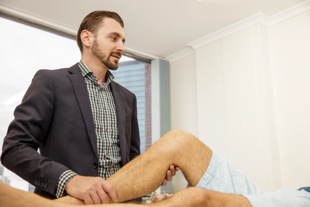
Patella stabilisation treats a kneecap that repeatedly slips or dislocates.
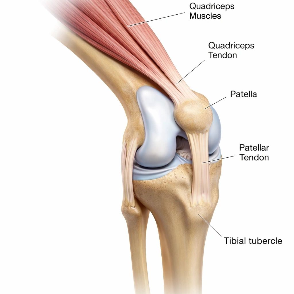
The patella (kneecap) normally sits within a groove at the front of the femur called the trochlea. As the knee bends and straightens, the patella moves within this groove.
Patellar instability occurs when the kneecap moves too far out of its normal position. This may involve:
- subluxation – the kneecap partially slips out
- dislocation – the kneecap completely leaves the groove
- repeated episodes of instability
- apprehension or fear that the kneecap is about to dislocate
The medial patellofemoral ligament (MPFL) is an important ligament on the inner side of the knee that helps prevent the patella moving excessively towards the outside. It is commonly injured when the kneecap dislocates.
However, patellar instability is not always simply a torn ligament. Other contributing factors can include:
- trochlear dysplasia – a shallow or unusually shaped groove
- patella alta – a relatively high kneecap
- increased lateral pull through the patellar tendon
- rotational or alignment differences in the leg
- generalised ligament laxity
A systematic review and meta-analysis of recurrence after first-time patellar dislocation found that younger age, open growth plates, trochlear dysplasia, patella alta and increased tibial tubercle–trochlear groove distance were associated with recurrent instability.
For this reason, patella stabilisation is not one operation. Surgery should address the particular factors causing instability in the individual knee.
Why is Patella Stabilisation Surgery Done?
Surgery is mainly considered for recurrent or ongoing symptomatic kneecap instability.
Not every patellar dislocation requires surgery.
A first-time dislocation can often be treated with physiotherapy, progressive strengthening, activity modification and sometimes temporary bracing.
Surgery may be considered when there is:
- recurrent patellar dislocation
- repeated subluxation or giving way
- persistent instability despite appropriate physiotherapy
- significant apprehension limiting sport or everyday activity
- an associated cartilage or osteochondral injury
- anatomical factors creating substantial ongoing instability
A systematic review and network meta-analysis comparing non-operative treatment, MPFL repair and MPFL reconstruction found lower redislocation rates after MPFL reconstruction than after non-operative treatment or MPFL repair, while also recognising that surgery introduces potential complications.
Even after a first dislocation, recurrence risk varies considerably. The systematic review of recurrence risk factors reported an overall recurrence rate of approximately 34%, but recurrence was substantially higher when several anatomical risk factors were present.
The decision to operate should therefore consider how often the kneecap is unstable, symptoms, anatomy, cartilage damage, activity requirements and response to rehabilitation, rather than simply the appearance of an MRI scan.
How is Patella Stabilisation Surgery Performed?
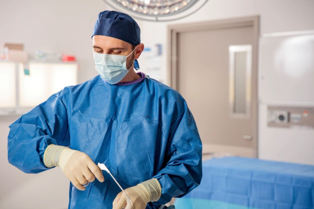
Surgery is tailored to the damaged ligament, kneecap alignment and shape of the knee.
The exact operation depends on why the patella is unstable. Some patients require only a ligament reconstruction, while others benefit from a combination of soft-tissue and bony procedures.
Anaesthetic
Patella stabilisation surgery is usually performed under a general anaesthetic, with additional local anaesthetic or regional techniques used for pain relief where appropriate.
Assessing the knee
An arthroscopy, or keyhole examination, may be performed to assess:
- cartilage on the patella
- cartilage within the trochlea
- loose cartilage or bone fragments
- menisci
- other structures inside the knee
Cartilage or osteochondral injuries can sometimes be treated during the same operation.
MPFL reconstruction
Medial patellofemoral ligament reconstruction is commonly used for recurrent lateral patellar instability.
A tendon graft is used to recreate the damaged MPFL between the inner side of the patella and the femur.
The aim is not to tightly hold the kneecap in one position. The graft is positioned and tensioned to provide stability while allowing the patella to move naturally within the trochlea as the knee bends.
A systematic review and meta-analysis of return to sport following MPFL reconstruction involving 930 patients reported a recurrent instability rate of approximately 2% across the included studies.
Tibial tubercle osteotomy
The tibial tubercle is the bony attachment of the patellar tendon on the upper shin bone.
In some patients, its position contributes to abnormal lateral pull on the kneecap or a high-riding patella.
A tibial tubercle osteotomy (TTO) involves carefully cutting this piece of bone, moving it to a more appropriate position and fixing it with screws while it heals.
Depending on the problem, the tubercle can be moved:
- towards the inside of the knee to alter the direction of pull
- forwards to reduce pressure on selected damaged cartilage
- downwards in selected patients with significant patella alta
Not everyone undergoing MPFL reconstruction needs an osteotomy.
A systematic review and meta-analysis comparing isolated MPFL reconstruction with MPFL reconstruction plus tibial tubercle osteotomy found similar functional outcomes and low redislocation rates with both strategies. Importantly, patients undergoing the combined procedure generally had different anatomical characteristics, highlighting the need to select the procedure according to the individual knee.
Trochleoplasty
Some patients have severe trochlear dysplasia, where the groove in which the kneecap should travel is very shallow or abnormally shaped.
In selected patients with severe dysplasia and recurrent instability, trochleoplasty may be considered to reshape the trochlea.
This is a specialised procedure and is not required for most patients.
A 2025 systematic review and meta-analysis of patellar stabilisation in patients with trochlear dysplasia found improved outcomes and low redislocation rates following MPFL reconstruction, trochleoplasty and combined procedures, but did not demonstrate that one operation was universally appropriate.
Closing the knee
Once the reconstruction is complete, the deeper tissues and skin are closed and dressings are applied.
A knee brace may be used depending on the procedure performed.
How Should I Prepare for Patella Stabilisation in Brisbane?

Preparing your knee, health and home can make early rehabilitation easier.
Prehabilitation
Where possible, physiotherapy before surgery aims to:
- reduce swelling
- restore full knee extension
- maintain knee flexion
- strengthen the quadriceps
- strengthen the hip and gluteal muscles
- improve balance and movement control
The aim is to arrive at surgery with the knee as mobile, strong and settled as reasonably possible.
Health preparation
Before surgery:
- stop smoking or vaping if possible
- maintain good nutrition
- optimise diabetes and other medical conditions
- provide a complete list of medications and supplements
- tell us about blood-thinning medications
- tell us about previous blood clots or anaesthetic problems
Preparing your home
You will generally require crutches during early recovery, and some patients also require a brace.
Arrange transport and some assistance with shopping, meals and everyday activities if required. Remove loose rugs and other trip hazards and consider how you will manage stairs.
Because Brisbane has a warm and humid climate, particularly during summer, consider making sure you have access to a cool, comfortable and preferably air-conditioned environment during early recovery. Apart from making recovery more comfortable, published research has found an association between warmer weather and higher rates of surgical-site infection.
Keeping yourself comfortable and your surgical dressing clean and dry is therefore particularly important during Brisbane's warmer months.
What Should I Expect After Patella Stabilisation Surgery?

Recovery progresses from protecting the reconstruction to rebuilding strength and confidence.
Rehabilitation depends on the operation performed. Recovery after an isolated MPFL reconstruction is different from recovery after MPFL reconstruction combined with an osteotomy or cartilage procedure.
Day surgery or hospital stay
Some patients can go home on the day of surgery, while others stay overnight.
Pain is managed using several different approaches. A physiotherapist will help you begin moving safely and teach you how to use crutches.
First 2 weeks
Early priorities include:
- controlling pain and swelling
- protecting the reconstruction
- regaining appropriate knee movement
- activating the quadriceps
- keeping the wounds clean and dry
- walking safely with crutches
Weight-bearing and knee-bending instructions depend on the operation performed.
2–6 weeks
Physiotherapy progressively focuses on knee movement, quadriceps activation and walking.
Patients who have undergone a tibial tubercle osteotomy generally require additional protection while the bone heals compared with patients having an isolated soft-tissue reconstruction.
A systematic review of rehabilitation after MPFL reconstruction with and without tibial tubercle osteotomy found considerable variation between published protocols and no single rehabilitation timetable suitable for every reconstruction.
6 weeks–3 months
As healing progresses, rehabilitation increasingly focuses on:
- restoring knee movement
- quadriceps strength
- hip and gluteal strength
- balance
- single-leg control
- normal walking
- cardiovascular fitness
Driving can resume when you can safely control the vehicle, are no longer impaired by pain medication and can confidently perform an emergency stop.
3–12 months
Running, jumping, pivoting and sport-specific exercises are progressively introduced once sufficient healing, strength and movement control have returned.
Return to sport should consider strength, stability, movement quality, confidence and the particular operation performed, rather than simply reaching a particular date.
What Are the Long-Term Outcomes After Patella Stabilisation?

Most patients improve, but recurrence and ongoing kneecap symptoms remain possible.
The primary aim of patella stabilisation surgery is to reduce recurrent instability and allow more confident use of the knee.
A systematic review and meta-analysis of MPFL reconstruction and return to sport found that approximately 93% of patients returned to some form of sport, while approximately 71% returned to or exceeded their previous level. Mean return was around 6–7 months.
A separate systematic review of return to play after MPFL reconstruction reported an overall return-to-play rate of approximately 85%, with around 68% returning to their previous level and recurrent instability occurring in approximately 5% of patients.
These figures describe groups of patients and cannot predict the outcome for an individual patient.
Some patients can continue to experience pain at the front of the knee despite improved stability, particularly when cartilage damage was already present.
Does patella stabilisation prevent arthritis?
This has not been established.
Patellar dislocation itself can injure the cartilage. A long-term MRI study following traumatic patellar dislocation found progressive patellofemoral cartilage deterioration over approximately eight years.
A larger population-based study of patellofemoral arthritis after lateral patellar dislocation also found a substantially increased long-term risk of clinically significant patellofemoral arthritis compared with people who had not sustained a dislocation.
However, this does not prove that stabilisation surgery prevents future arthritis.
The primary purpose of surgery is to treat symptomatic instability and address relevant anatomical factors.
Warning Signs After Patella Stabilisation
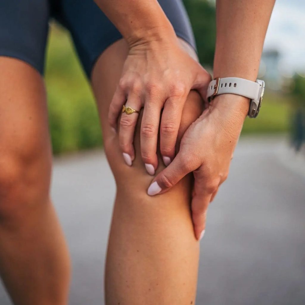
Worsening wounds, severe swelling, instability or breathing symptoms require assessment.
Contact our office, your GP or seek medical attention if you develop:
Possible infection
- increasing redness or warmth around the wounds
- worsening pain after initially improving
- persistent wound leakage or pus
- fever or feeling significantly unwell
Possible blood clot
Seek medical attention for new calf pain or significant one-sided leg swelling.
Sudden shortness of breath, chest pain or coughing blood requires emergency assessment.
Increasing stiffness
Some stiffness is expected during early recovery.
Progressive loss of movement or difficulty regaining knee flexion or extension should be discussed with your treating team.
Wound, nerve or circulation problems
Persistent wound leakage, wound opening, rapidly increasing swelling, a cold or pale foot, or new significant weakness requires assessment.
Recurrent instability
Some apprehension around the kneecap can occur while strength and confidence recover.
However, a definite episode where the kneecap significantly slips or dislocates should be assessed.
New injury
A significant fall, twist or sporting injury can potentially damage a healing reconstruction or osteotomy.
New severe pain, swelling, instability or loss of function should be reviewed.
Procedures explained
Patella stabilisation

Dr Alexey Borshch is a specialist orthopaedic surgeon in Brisbane with a particular clinical focus on surgery of the hip, knee and lower limb.
His practice as a Brisbane orthopedic surgeon focuses on the assessment and treatment of knee injuries, as well as performing ACL reconstruction surgery, PCL reconstruction, Multi-ligament reconstruction and patella stabilisation surgery.
Dr. Borshch completed his medical degree at the University of Queensland before undertaking specialist orthopaedic training in Queensland. He completed the Australian Orthopaedic Association training program and the Royal Australasian College of Surgeons Fellowship Examination in Orthopaedic Surgery. He obtained specialist registration in Orthopaedic Surgery in 2022 and is a Fellow of the Australian Orthopaedic Association.
He offers consultations in Hamilton, Brisbane, and conducts private orthopaedic surgery at Brisbane Private Hospital.